444 4 484
info@gucluhanguclu.com
Beşyol, Florya, Akasya St. No:4 Apt:1, 34295 Küçükçekmece/Istanbul
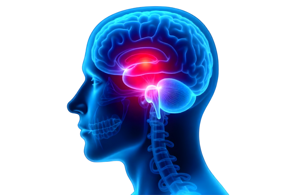
The pituitary gland is known as the central control organ of the endocrine system and plays critical roles in the body. Located at the base of the brain, just beneath the hypothalamus and behind the nasal cavity, the pituitary gland is about the size of a pea. However, through the hormones it secretes, it affects nearly every function of the body. It directly influences vital functions such as body growth, metabolic rate, reproduction, stress responses, and water balance. For this reason, the pituitary gland is often referred to as the "master gland."
Anatomically, the pituitary gland consists of two main lobes: the anterior pituitary (adenohypophysis) and the posterior pituitary (neurohypophysis). Both lobes have different types of cells and secrete distinct hormones. These hormones play a critical role in maintaining homeostasis by sending signals that affect various organs and systems in the body.

The anterior pituitary produces hormones that regulate most of the body's essential physiological functions. Signals from the hypothalamus stimulate hormone secretion from the anterior pituitary. The main hormones secreted by the anterior pituitary include:
The posterior pituitary is controlled by nerve signals from the hypothalamus and directly secretes two important hormones:
The pituitary gland works in close collaboration with the hypothalamus. Located in the brainstem, the hypothalamus sends signals to the pituitary gland to start or stop hormone production. This feedback loop between the hypothalamus and the pituitary gland forms a critical system for maintaining hormonal balance. When specific hormone levels rise or fall, the hypothalamus adjusts the activity of the pituitary gland accordingly.
This hormonal control mechanism allows the body to adapt to internal and external environmental changes while maintaining homeostasis. For example, when the body loses water, ADH secretion increases to promote water reabsorption. Similarly, in stressful situations, the pituitary gland increases ACTH secretion, prompting the adrenal glands to produce cortisol.
Hormones produced by the pituitary gland affect nearly every organ and system in the body. Any dysfunction in this gland can disrupt the balance of many bodily systems. For example, a tumor in the pituitary gland can either excessively increase hormone production (leading to conditions like prolactinoma or acromegaly) or suppress it. Consequently, any disorder in the pituitary gland can result in significant health issues due to hormonal imbalances and neural pressure.
In summary, the pituitary gland plays a crucial role in regulating vital bodily functions. This small gland is responsible for secreting hormones that control processes like growth, reproduction, metabolism, stress response, and water balance. Given the wide-ranging health issues that can arise from pituitary dysfunction, maintaining the gland's healthy operation is of utmost importance.
A pituitary gland tumor is a mass formed by the uncontrolled and abnormal growth of cells in the pituitary gland. Pituitary tumors are typically classified as benign (non-cancerous), meaning they do not tend to spread to surrounding tissues. However, this does not mean that pituitary tumors are harmless. Due to their location and their effect on hormone production, these tumors can lead to significant health problems.
The effects of pituitary tumors depend on their size and functional characteristics. As the pituitary gland is a central hub for the body's hormonal system, tumors in this gland can directly impact hormone production and create pressure on surrounding nerves as they grow.
Pituitary tumors are generally categorized into two main types: functional (hormone-producing) tumors and non-functional (non-hormone-producing) tumors. This distinction is based on whether the tumor produces hormones and how this affects the body.
Functional pituitary tumors produce an excess of hormones. These tumors can cause significant symptoms due to hormonal imbalances, even when they are small in size. Since the pituitary gland produces various hormones, the health issues that arise depend on which hormone is being overproduced:
Non-functional pituitary tumors do not produce hormones. These tumors cause problems when they grow and begin to press on surrounding tissues. Non-functional tumors typically present symptoms based on their size, often compressing the optic nerves and causing vision problems. Other symptoms may include severe headaches, hormonal deficiencies (hypopituitarism), and fatigue.
Pituitary tumors are also classified based on their size:
While pituitary tumors do not directly spread to brain tissue, they can exert pressure on surrounding tissues, leading to severe symptoms. For instance, when tumors press on the optic chiasm near the pituitary gland, vision loss is one of the most commonly observed effects. Additionally, depending on the tumor’s size, headaches, hormonal imbalances, and neurological symptoms may also occur.
Pituitary tumors can disrupt the body’s overall balance by either increasing or suppressing hormone production. Excessive or insufficient hormone levels can affect systems such as metabolism, reproduction, stress response, and growth over the long term. If left untreated, these conditions can significantly reduce patients’ quality of life and even lead to life-threatening complications. Early diagnosis and treatment of pituitary gland tumors are therefore critically important.
The exact causes of pituitary gland tumors are not fully understood, but genetic and environmental factors are thought to contribute to their development. Pituitary tumors often arise spontaneously without a clear cause. However, certain individuals may have an increased risk due to genetic predispositions and external factors.
Some pituitary tumors are associated with genetic factors. In such cases, genetic mutations or hereditary syndromes can lead to abnormal growth of pituitary cells. Genetic factors in pituitary tumors are commonly linked to the following syndromes:
In addition to genetic causes, environmental factors may play a role in the development of pituitary tumors. External influences, in particular, are thought to contribute to pituitary tumor formation by affecting cell growth and hormone production.
Beyond specific genetic and environmental causes, there are several risk factors that may contribute to the development of pituitary tumors. While these factors may not directly trigger tumor formation, they can increase an individual’s susceptibility:
Hormonal disorders play a significant role in the development of pituitary tumors. As one of the most critical glands regulating hormonal balance in the body, disruptions in pituitary hormone production can lead to excessive cell proliferation. The following conditions, in particular, are associated with an increased risk of pituitary tumors:
Although pituitary tumors are relatively rare in the general population, autopsy studies indicate that up to 10% of individuals may have pituitary tumors. However, most of these tumors are very small and do not cause noticeable clinical symptoms, remaining undetected throughout a person's lifetime. While the majority of pituitary tumors are benign and not cancerous, their growth or hormonal imbalances can lead to significant symptoms.
The causes and risk factors of pituitary tumors highlight the critical role of genetic predispositions, environmental influences, and hormonal imbalances in tumor development. Therefore, early diagnosis and regular monitoring are essential for effectively managing pituitary tumors.
The symptoms of pituitary gland tumors vary widely depending on the tumor’s size, location, and whether it is functional or non-functional. Some of these tumors cause hormonal imbalances, while others exert pressure on surrounding structures as they grow. Pituitary gland tumors generally grow slowly, meaning symptoms often progress over time.
One of the most common symptoms of a pituitary tumor is headaches. As the tumor grows, it can exert pressure on the tissues surrounding the pituitary gland. This pressure may cause a sensation of tightness or a persistent ache, often localized in the front of the head. While headaches are one of the initial signs of pituitary tumors, they are not unique to this condition and can be mistaken for other causes. Careful evaluation is necessary to distinguish them from other headache types.
Since the pituitary gland is located just below the optic nerves (optic chiasm), a growing tumor can compress these nerves, leading to visual impairments. As the tumor enlarges or affects the nerves controlling eye movement and visual fields, the following vision problems may occur:
The pituitary gland regulates many hormonal balances in the body, and tumors in this gland can disrupt hormone production. These disruptions may lead to excessive or insufficient production of certain hormones, causing a wide variety of symptoms depending on the specific hormones involved.
Functional pituitary tumors are associated with symptoms resulting from overproduction of hormones. On the other hand, non-functional tumors cause symptoms as they grow and compress surrounding nerves and tissues.
Functional pituitary tumors secrete excess hormones, disrupting hormonal balance and leading to various clinical syndromes:
Non-functional pituitary tumors do not secrete hormones but can cause symptoms by pressing on surrounding nerves and tissues as they grow. Common symptoms include headaches, visual impairments, and suppressed hormone production by the pituitary gland (hypopituitarism):
Additional symptoms of pituitary tumors vary based on the tumor’s size and location. Large pituitary tumors (macroadenomas) can compress surrounding tissues and cause neurological symptoms:
Pituitary gland tumors present a wide range of symptoms depending on their size, hormonal activity, and effects on surrounding tissues. These symptoms can aid in early detection and facilitate the development of effective treatment strategies.
Diagnosing pituitary gland tumors is often a complex process that requires a multidisciplinary approach. During diagnosis, doctors evaluate the patient’s symptoms and use various imaging methods, hormone tests, and neurological assessments to gather detailed information about the tumor’s size, location, and functionality. Here are the primary methods used in diagnosing pituitary tumors:
MRI is the most commonly used imaging method for diagnosing pituitary gland tumors. It provides detailed images of the pituitary gland and surrounding tissues, allowing evaluation of the tumor's size, location, and impact on adjacent structures. MRI is particularly effective for detecting small tumors (microadenomas). It is also used to determine whether the tumor is pressing on the optic nerves and whether surgical intervention is necessary. MRI helps precisely map the tumor and supports the creation of an optimal treatment plan for the patient.
While not as detailed as MRI, CT scans may be used in certain situations. CT scans are preferred in cases where MRI is not accessible or cannot be performed (e.g., in patients with pacemakers). CT scans are used to evaluate calcifications within the pituitary tumor or the effects of the tumor on surrounding bone structures. They are particularly helpful in identifying large pituitary tumors (macroadenomas) and aiding in surgical planning.
Hormone tests play a crucial role in determining whether the pituitary gland tumor is related to abnormal hormone production. Since the pituitary gland regulates the production of various hormones, blood tests are conducted to assess whether the tumor is causing an increase in hormone levels. These tests help identify if the tumor is functional and which hormones are affected. Hormone tests are used for the following evaluations:
Pituitary gland tumors can compress the optic nerves, leading to vision problems. For this reason, visual field tests are often conducted in patients suspected of having pituitary tumors. If the tumor is large and pressing on the optic nerves, the patient may experience narrowing of the visual field. The most common visual impairment seen in pituitary tumors is bitemporal hemianopia, characterized by loss of peripheral vision in both eyes. This symptom indicates that the tumor is compressing the optic nerves, causing visual field defects.
Pituitary tumors can cause not only hormonal imbalances but also a range of neurological symptoms. For this reason, comprehensive neurological evaluations are performed in patients suspected of having pituitary tumors. Assessments of headaches, visual impairments, fatigue, weakness, and neurological reflexes help determine the tumor's impact on the central nervous system.
In some cases, specialized tests may be used to diagnose pituitary tumors. For instance, Dynamic Endocrine Testing is conducted to evaluate the pituitary gland’s ability to produce specific hormones. These tests involve administering certain hormones to the patient and measuring how the body responds. These tests are particularly important in assessing whether hormone levels are regulated in cases of functional pituitary tumors.
Diagnosing pituitary tumors is the first step in planning treatment. The appropriate treatment method is determined based on the tumor’s size, hormone production, and the patient’s overall health status. Monitoring and follow-up may be sufficient for small and non-functional tumors, whereas large or hormone-producing tumors may require surgical intervention, radiation therapy, or medication.
Treatment methods for pituitary tumors vary depending on the tumor’s size, the patient’s overall health, and whether the tumor produces hormones. Treatment options range from surgical interventions to medication, with each case requiring a personalized approach. Below are the main treatment methods used for pituitary gland tumors:
Surgical intervention is the most commonly chosen treatment method for pituitary gland tumors, especially in cases involving large tumors or symptoms caused by hormone production. While pituitary tumors are generally benign, their size or hormone production can exert pressure on surrounding tissues, leading to serious complications. Therefore, surgery plays a critical role in relieving tumor pressure and balancing hormone levels.
Transsphenoidal surgery is the most common and minimally invasive method for removing pituitary tumors. The surgeon accesses the pituitary gland through the nostrils or the inner part of the upper lip to remove the tumor. This technique is preferred for small or medium-sized pituitary tumors. Below are the advantages and stages of this method:
Craniotomy is a more extensive and invasive method used to surgically remove pituitary tumors. It is generally preferred when transsphenoidal surgery is not feasible. This approach is used for large tumors or those that have spread deep into the pituitary gland. The craniotomy procedure involves temporarily removing a portion of the skull to directly access the tumor.
The success rate of surgical intervention depends on factors such as the tumor’s size, whether it produces hormones, and the surgeon’s expertise. For small and non-functional tumors, surgical success rates are very high, and most patients experience a return to normal hormone levels after surgery. Functional tumors may also show rapid normalization of hormone levels, leading to a reduction in symptoms.
However, in some cases, the entire tumor cannot be removed, or the tumor may regrow. For this reason, regular follow-up and imaging are essential after surgery. The likelihood of tumor recurrence is higher in functional tumors, which may necessitate additional treatments.
Regular monitoring of patients after surgical intervention is critical. MRI scans are conducted regularly to check for tumor regrowth. Additionally, hormone levels are monitored to confirm normalization. In some cases, hormone replacement therapy may be necessary after surgery. If the pituitary gland has been damaged, reduced hormone production may require lifelong hormone therapy.
The follow-up process after surgery includes long-term monitoring to mitigate the risk of recurrence. This process is essential for maintaining the patient’s quality of life and ensuring proper functioning of the pituitary gland.
Radiation therapy is used to prevent tumor regrowth after surgery or to eliminate tumor cells that could not be completely removed surgically. It can be an effective treatment option, especially in cases where complete surgical removal is not possible or surgery poses a high risk. There are two main types of radiation therapy:
One disadvantage of radiation therapy is that its effects may take time to manifest. Stopping tumor growth and alleviating symptoms can take several months. Additionally, some patients may develop damage to other parts of the pituitary gland, which may necessitate hormone replacement therapy.
Medication therapy plays a significant role, particularly in hormone-producing pituitary tumors. Functional tumors secrete excessive amounts of hormones, causing hormonal imbalances, and medication therapy aims to correct these imbalances. Various drugs are used to reduce hormone production or slow tumor growth:
Medication therapy can also be used preoperatively to shrink the tumor in some patients. Additionally, it can be effective in cases where surgery cannot be performed or where the tumor recurs.
Some pituitary tumors are small, do not produce hormones, and do not pose significant harm to the patient. These tumors are typically monitored routinely with a 'watchful waiting' approach. During this process, regular MRI scans and hormone tests are conducted, and treatment options are reassessed if the tumor grows or symptoms develop. This strategy is often preferred in older patients or those with high surgical risks.
During the follow-up process, patients are closely monitored for symptoms, and if tumor growth or hormonal imbalances are detected, the treatment plan is re-evaluated. This approach is suitable when surgical intervention or radiation therapy is unnecessary and poses fewer risks to the patient.
In some cases, combination therapy may be required. Radiation therapy or medication therapy may be added following surgical intervention. These combinations are particularly effective when large tumors cannot be completely removed surgically or when hormone production cannot be adequately controlled.
The postoperative recovery process for pituitary gland tumor surgery varies depending on the patient’s overall health, the size of the tumor, and the type of tumor removed. The type of surgical procedure, whether minimally invasive or more extensive, also impacts recovery. Careful recovery and follow-up are essential to restoring the patient’s quality of life and preventing potential complications.
Most patients are observed in the hospital for a few days after surgery. For minimally invasive methods such as transsphenoidal surgery, patients can typically be discharged within 2-3 days, while more extensive procedures like craniotomy may require a longer hospital stay. During this period, patients are closely monitored, vital signs are checked, and potential postoperative complications are identified early.
One of the most common complications in the postoperative period is infection. In pituitary gland surgeries, especially those performed through the nasal route, there is a risk of infection in surrounding tissues. Additionally, skin infections resulting from the surgery may also occur.
Since the pituitary gland is responsible for producing various hormones, hormonal balance can be a significant concern after surgery. Parts of the pituitary gland may be damaged or completely removed during surgery, leading to hormonal imbalances.
Despite surgical removal of pituitary tumors, regrowth may occur in some cases. The likelihood of recurrence depends on factors such as the tumor type, size, and the extent of removal during surgery. Therefore, regular follow-up is crucial to monitor the risk of recurrence.
Some potential complications after surgical treatment of pituitary gland tumors include:
In the postoperative period, patients require careful monitoring. Even if surgical intervention is successful, patients may need lifelong regular follow-ups. Long-term management of disruptions in the pituitary gland’s hormone production is essential, and continuous monitoring of hormonal balances is critical to improve patients’ quality of life.
If left untreated, pituitary tumors can grow over time and interfere with hormone production, leading to serious complications. The long-term problems caused by untreated pituitary tumors vary depending on the tumor’s size, location, and whether it produces hormones. These complications are often progressive and permanent, making early diagnosis and appropriate treatment of pituitary tumors critically important.
The pituitary gland is located just below the brain and near the optic nerves. As pituitary tumors grow, the likelihood of them pressing against the optic nerves increases. This pressure can gradually lead to vision impairments and, if untreated, permanent vision loss.
Functional pituitary tumors can produce excessive hormones, disrupting the body’s hormonal balance. If untreated, these tumors can cause severe hormonal imbalances and associated diseases over the long term.
Growing pituitary tumors can put pressure not only on the optic nerves but also on surrounding brain tissues and nerves, leading to various neurological problems.
Pituitary tumors that disrupt hormone production can lead to general metabolic dysfunction in the body. For example, tumors that produce thyroid-stimulating hormone (TSH) can cause hyperthyroidism (overactive thyroid) or hypothyroidism (underactive thyroid), which can affect metabolism and result in serious health problems.
Untreated pituitary tumors can have significant effects not only on physical health but also on the overall quality of life.
After pituitary tumor surgery or treatment, long-term care and follow-up are vital. The post-treatment period must be carefully managed, considering factors such as the risk of tumor recurrence, hormonal imbalances, and effects on quality of life.
Removing part of the pituitary gland during surgery can lead to a reduction in hormone production, necessitating lifelong hormone replacement therapy. This therapy may include supplementation of thyroid hormones, growth hormones, cortisol, or sex hormones to address deficiencies.
During the post-treatment period, regular MRI or CT scans are performed to check for tumor regrowth. The hormone production of functional tumors is also monitored. Given the risk of tumor recurrence after surgery, these imaging studies typically continue lifelong.
Adopting a healthy lifestyle after pituitary tumor treatment can accelerate recovery and support overall health.
Untreated pituitary tumors can lead to vision loss, hormonal imbalances, neurological problems, and long-term severe health issues. Early diagnosis and treatment are crucial to prevent these complications and maintain patients’ quality of life.

